

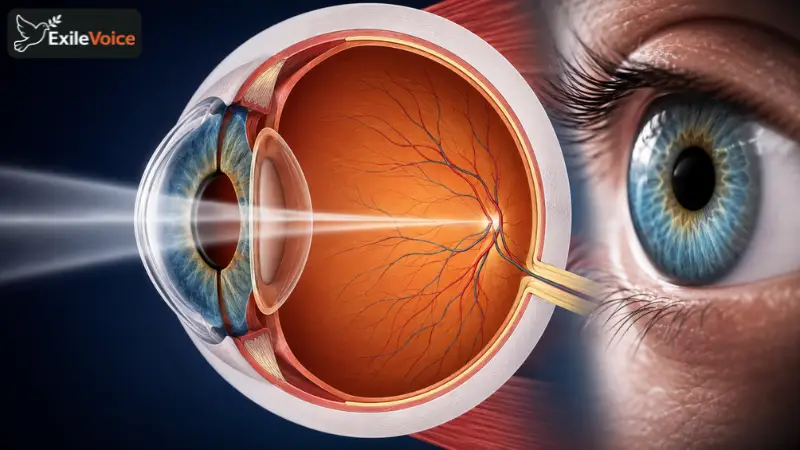
The human eye is one of the most sophisticated and intricate biological systems within the human body, operating as a high precision optical instrument that translates light waves into coherent visual imagery. Understanding the anatomical mechanics of the eye reveals how seamlessly biological structures collaborate to perceive the surrounding world. Light is the fundamental catalyst for sight, reflecting off objects in our environment and entering the optical pathway through the anterior structures of the eye.
The journey of light begins at the cornea, a transparent, dome shaped protective window that covers the front portion of the eye. The cornea performs the initial and significant refraction of incoming light rays, bending them so they can pass efficiently through the subsequent layers. Positioned immediately behind the cornea is the anterior chamber, a specialized space filled with aqueous humor, a clear fluid that nourishes surrounding tissues and maintains internal ocular pressure.
As light continues its path, it encounters the iris, the colored ring of muscle tissue that gives eyes their distinct hue. Centered within the iris is the pupil, a dynamic aperture that regulates the volume of light entering the inner eye. Through the coordinated contraction and relaxation of muscular fibers within the iris, the pupil dilates in dim environments to maximize light capture and constricts in bright settings to protect the delicate internal receptors from overexposure.
Directly behind the iris lies the crystalline lens, a flexible, transparent structure that performs secondary refraction. The lens dynamically fine tunes its focal length through a process called accommodation, allowing the eye to shift focus seamlessly between distant horizons and close up objects. Once the light rays pass through the lens, they traverse the vitreous humor and project onto the retina, a highly specialized light sensitive tissue lining the interior back wall of the eye.
The retina houses millions of photoreceptor cells known as rods and cones. Rods are exceptionally sensitive to low light levels and motion, enabling night and peripheral vision, whereas cones are responsible for high resolution color vision and fine detail under bright illumination. Upon capturing photons, these photoreceptors convert optical signals into electrochemical impulses. These nerve signals travel along the optic nerve directly to the occipital lobe of the brain, which decodes the data, corrects image inversion, and constructs our final conscious visual perspective.
Variations in the physical geometry of the eyeball frequently lead to common refractive errors. For instance, a myopic or nearsighted eye possesses an elongated axial length, causing light rays to converge in front of the retina rather than precisely upon it, which blurs distant objects. Conversely, hyperopia or farsightedness occurs when the eyeball is relatively short, forcing the focal point behind the retina and impairing near vision. Modern optometry successfully corrects these anatomical discrepancies using tailored concave or convex corrective lenses that restore the accurate focal point.